


2021 Vol. 7, No. 4

Cover Story
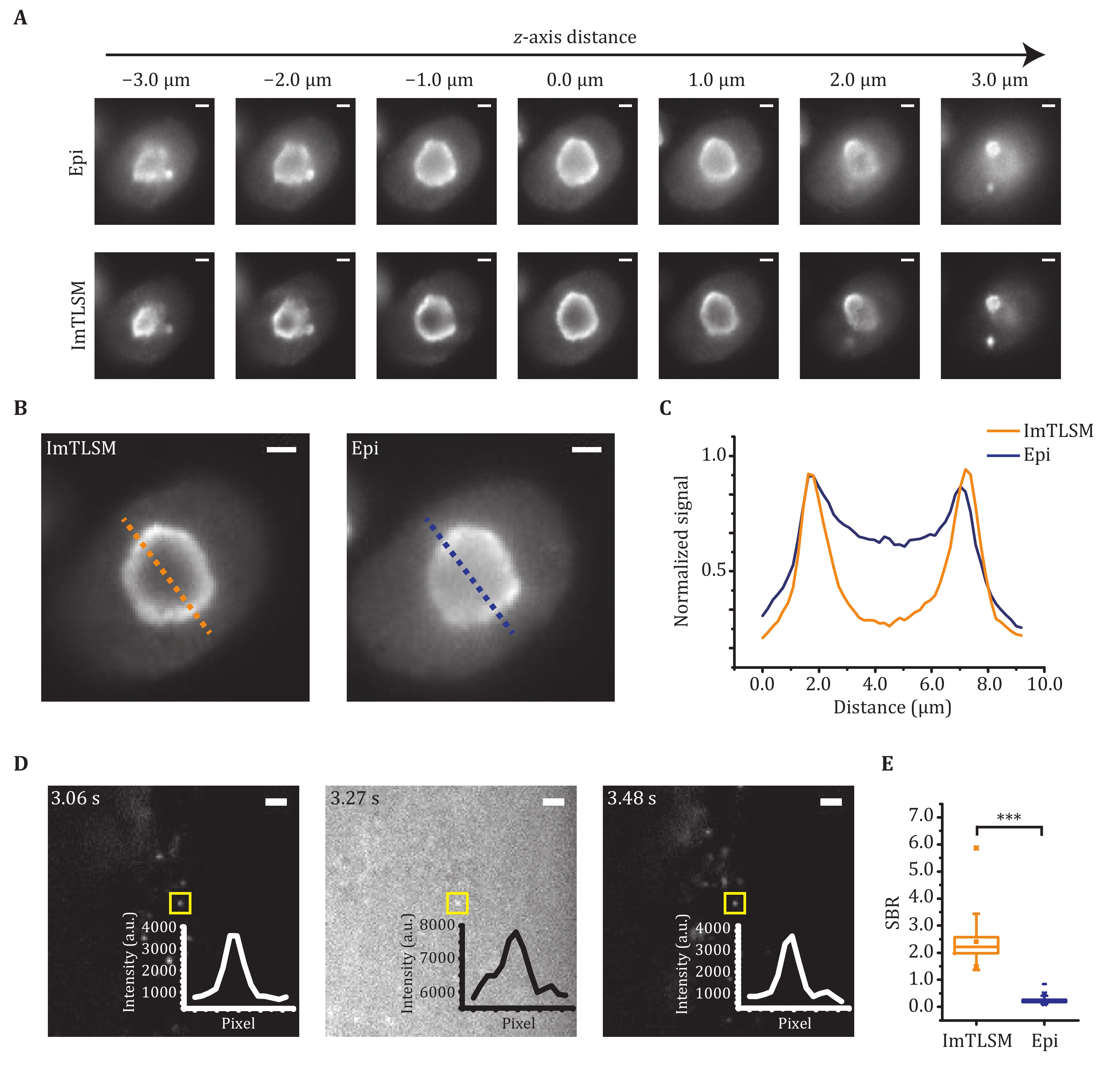
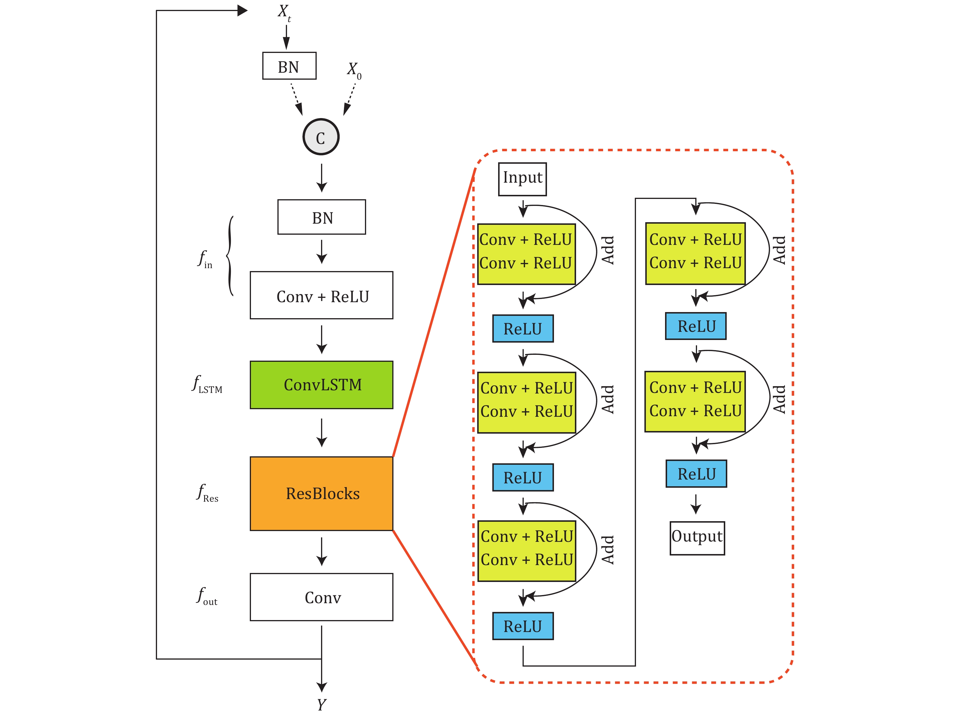
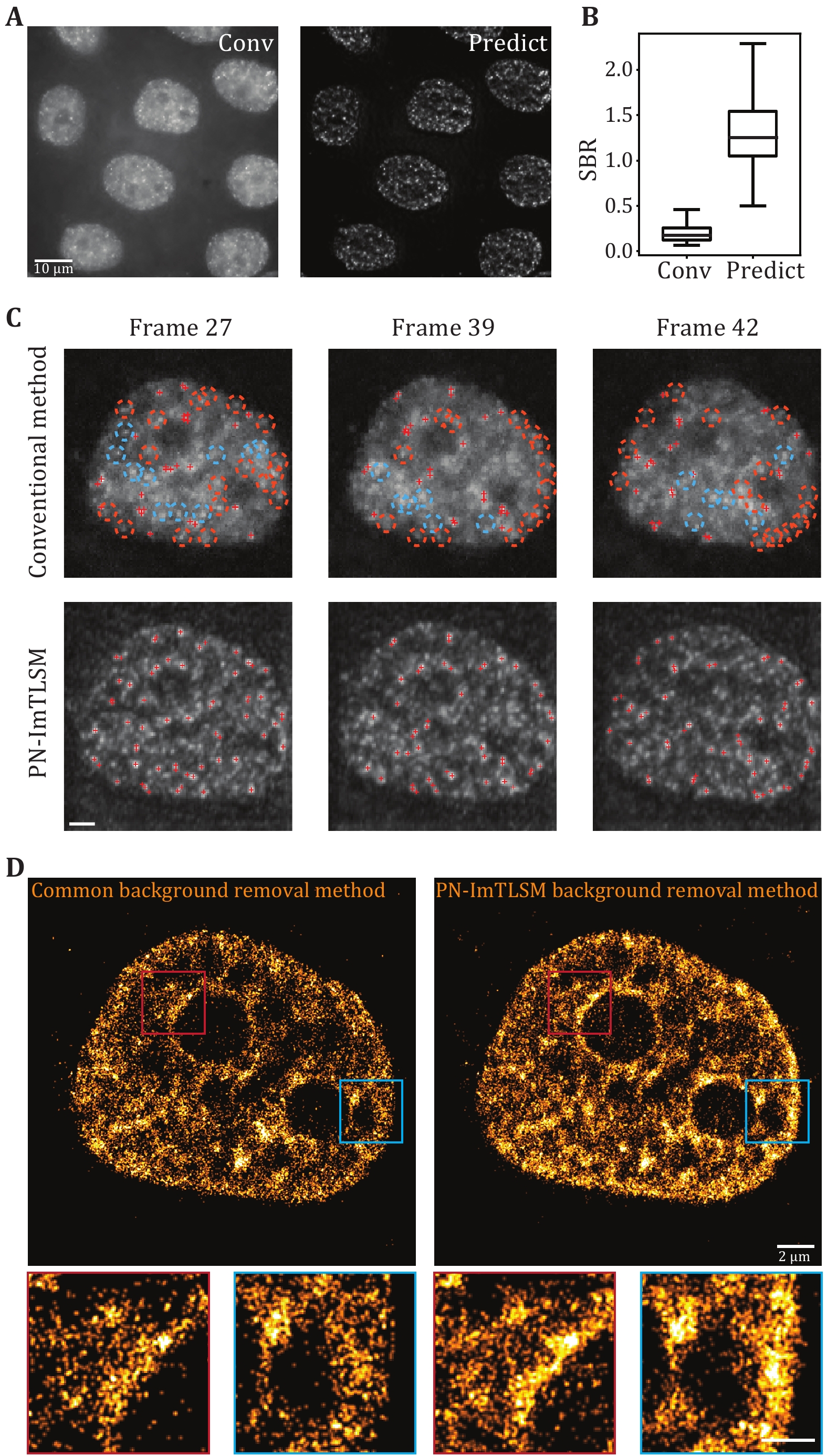
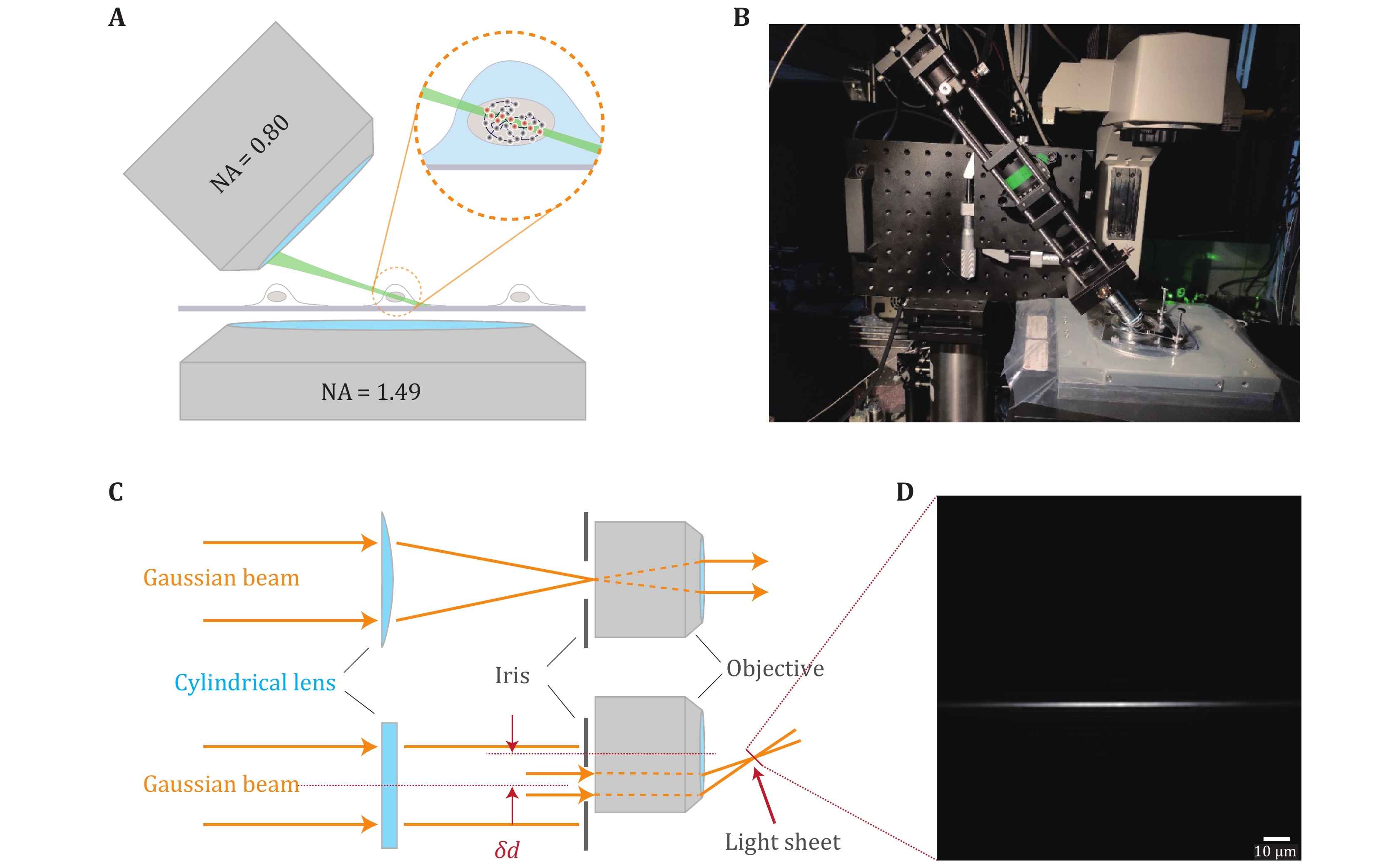
When imaging the nucleus structure of a cell, the out-of-focus fluorescence acts as background and hinders the detection of weak signals. Light-sheet fluorescence microscopy (LSFM) is a wide-field imaging approach which has the best of both background removal and imaging speed. However, the commonly adopted orthogonal excitation/detection scheme is hard to be applied to single cell imaging due to steric hindrance. For LSFMs capable of high spatiotemporal single cell imaging, the complex instrument design and operation largely limit their throughput of data collection. Here, the authors propose an approach for high-throughput background-free fluorescence imaging of single cells facilitated by the Immersion Tilted Light Sheet Microscopy (ImTLSM). ImTLSM is based on a light-sheet projected off the optical axis of a water immersion objective. With the illumination objective and the detection objective placed opposingly, ImTLSM can rapidly patrol and optically section multiple individual cells while maintaining single-molecule detection sensitivity and resolution. Further, the simplicity and robustness of ImTLSM in operation and maintenance enables high-throughput image collection to establish background removal datasets for deep learning. Using a deep learning model to train the mapping from epi-illumination images to ImTLSM illumination images, namely PN-ImTLSM, the authors demonstrated cross-modality fluorescence imaging, transforming the epi-illumination image to approach the background removal performance obtained with ImTLSM. They demonstrated that PN-ImTLSM can be generalized to large-field homogeneous illumination imaging, thereby further improving the imaging throughput. In addition, compared to commonly used background removal methods, PN-ImTLSM showed much better performance for areas where the background intensity changes sharply in space, facilitating high-density single-molecule localization microscopy. In summary, PN-ImTLSM paves the way for background-free fluorescence imaging on ordinary inverted microscopes.

 Abstract
 PDF
PDF


 Submit Your Paper
 For Author
 Templates


 (86-10) 64888458


 E-mail alert
 RSS
RSS